descriptive studyDescriptive study includes activities relates to characterizing the distribution of disease within a population. Descriptive studies can yield valuable information about a population's health
status, and they can be used to measure risks and generate hypotheses. It is also useful in health service evaluation and can be used periodically to determine whether a particular service is improving

The type of descriptive study

  • Case reports
  • Case series
  • Cross sectional studies
  • Ecologic studies

Case reports and series

Case report: describes an observation in a single patient.
ª  “I had a patient with a cold who drank lots of orange juice and got better.  Therefore, orange juice may cure colds.”
Case series: same thing as a case report, only with more people in it.
ª  “I had 10 patients with a cold who drank orange juice….”

Cross sectional studies

A cross-sectional study is a descriptive study in which disease and exposure status are measured simultaneously in a given population. Also called a “survey” or “prevalence” study Cross-sectional studies can be thought of as providing a "snapshot" of the frequency and characteristics of a disease in a population at a particular point in time. This type of data can be used to assess the prevalence of acute or chronic conditions in a population.

Research aim of prevalence survey

  • To describe distribution of disease
  • To discovery clue of pathogenesis
  • Be used in secondary prevention
  • To evaluate prevention and cure effect
  • Surveillance of disease
  • Health demand, health project and health policy decision

Describe the distribution of disease or health status by person, place and time, then analyze that which factors are relate to the disease or health status.

Secondary prevention seeks to minimize adverse outcomes of disease through early detection, even before symptoms develop and care is sought. Mammography for early detection of breast cancer in asymptomatic women is an example.

An occupational physician planning a coronary prevention program might wish to know the prevalence of different risk factors in the workforce under his care so that he could tailor his intervention accordingly.


The acute phase response is a quickly mobilized non-specific defensive response elicited in the response of the host to infection, tissue injury or inflammation.

Acute phase proteins 

Acute phase proteins have been defined as the proteins whose plasma concentration increases (positive acute phase proteins) or decreases (negative acute phase proteins) during the acute phase responses.

Acute phase proteins: Composition and Resource

APs are synthesized mainly by hepatic cells, the monocytes, and fibroblasts.

Acute phase proteins: Main Functions

1. To help the immune system adapt to various environmental stresses. removal of exogenous substance and necrotic tissue to promote the healing .for example:c-reactive protein c-reactive protein + combine with bacterial wall activate complement classic pathway promote phagocytosis induce the expression of inflammatory cytokines and tissue factor in monocytes.
2. Inhibit the activity of protease: The protease inhibitors can protect tissues from over injuries by protease 
3. up-regulation of fibrinogen in the acute phase response is beneficial  to increasing the hemostatic power and the wound healing
4. ceruloplasmin, haptoglobin, and hemopexin can bind copper and heme, and avoid the injuries of tissues by over many copper and iron. Ceruloplasmin exhibits an antioxidant function against oxidative stress injuries.
5. serum amyloid protein A associated HDL3 seems to facilitate the uptake and removal of cholesterol from monocytes/macrophages at the inflammatory site


Material metabolism in the liver

liver metabolism
  • carbohydrate metabolism
  • lipid metabolism
  • protein metabolism
  • vitamin metabolism
  • hormone metabolism 

Carbohydrate metabolism

Functions

Maintaining the constancy of the concentration of sugar in blood; supplying energy to peripheral tissues, especially to brain and erythrocytes.

Main pathways for carbohydrate metabolism

  • gluconeogenesis
  • glycogenesis and glycogenolysis
  • glycolysis 

Carbohydrate metabolism in varied nutrient conditions in liver

    --  glycogenesis↑
          --  excess carbohydrate is converted to fatty acids and exported in VLDL form
  • postabsorptive state
     --  glycogenolysis↑→release glucose to replenish blood sugar
  • starvation state
     --  gluconeogenesis
     --  lipid mobilization↑→acetone body↑→saving glucose

Lipid metabolism


Functions: 

Play important role in digestion, absorption, synthesis, catabolism and transportation of lipids.  

 Main pathways lipid metabolism

  • oxidation of fatty acids
  • synthesis of fatty acids and esterification
  • formation of ketone bodies
  • synthesis of lipoproteins and apolipoproteins (VLDLHDLapoC II)
  • degradation of lipoproteins

Role of liver in varied processes of lipid metabolism

  • digestion and absorption
    --  biliation.  Bile acid is essential for lipid digestion and  absorption
     --  liver dysfunction→fatty diarrhea
  • synthesis
     --  fatty acid, triacylglycerol, ketone body, cholesterol, phospholipid
     --  liver dysfunction→fatty liver, Ch/ChE↑
  • catabolism
     --  β-oxidation of fatty acids, degradation and excretion of cholesterol, degradation of LDL
  • tranportation
     --  synthesis and secretion of VLDL, HDL, apoC II, and LCAT

Protein metabolism


Functions: synthesis (γglobulin is excepted), secretion and elimination (albumin is excepted) of plasma proteins.

Liver dysfunction:

  • albumin↓→edema, A/G↓
  • plasma thromboplastin component ↓ →disturbance of blood coagulation
  • blood ammonia
  • amine↑→hepatic encephalopathy 

Vitamin metabolism

Functions

  • absorption of fat-soluble vitamins
  • storage of vitamins (VA, VK,VE,VB12)
  • transportation of vitamins synthesis of retinol-binding protein and VitD-binding protein
  • transformation of vitamins
  • β-carotene→VitA Vit D3 → 25-(OH)-Vit D3 water-soluble vitamins →cofactors

Liver dysfunction

  • biliary obstruction→VitK↓→hemorrhagic tendency
  • hepatic rickets

Hormone metabolism

Functions: inactivation of hormones
Manner: biotransformation

Liver dysfunction:

  • Spider angioma of skin
  • liver pulms
  • cutaneous pigmentation
  • hypoglycemia


Magnesium function
Magnesium is the fourth most common cation in the human body and the second most abundant intracellular cation in the human body. The human body contains approximately 24g (1000mmole) of magnesium and it is involved in many intracellular processes and is very essential for life. Metabolic irregularity or disturbance is associated with various abnormalities. The main sources of magnesium are vegetables, soybeans, nuts, whole grain cereals, eggs, and seafood. The minimum daily dietary magnesium intake to maintain magnesium balance in a normal person is about 240mg per day.

Magnesium functions – Co-factor and agitator of many enzymes

Magnesium plays an important role in the functions of more than 300 enzymes, Na+. K+ ATPase, hexokinase, choline esterase. It actively participates in many metabolic activities in the body. Magnesium helps to metabolize proteins, lipids, carbohydrates, and nucleic acid. Magnesium is also vital in regulating the cellular distribution of sodium and potassium through involvement in Na+, K+ ATPase.

Magnesium functions -Maintains irritability of the cells

Magnesium acts as an inhibitor to the central nervous system, neuromuscular and cardiac muscles. For neuromuscular irritability magnesium and calcium are synergic and for cardiac muscles they are the antagonist.

Magnesium functions – Maintains homeostasis of cells

Magnesium is an essential cofactor in correlative enzymes of DNA, cell cycle and apoptosis. In plasma, magnesium is important for maintaining DNA structure and veracity of DNA replication and activating DNA repairing including nucleotide excision repair, base excision repair, miss match repair, and microtubule assembly. 


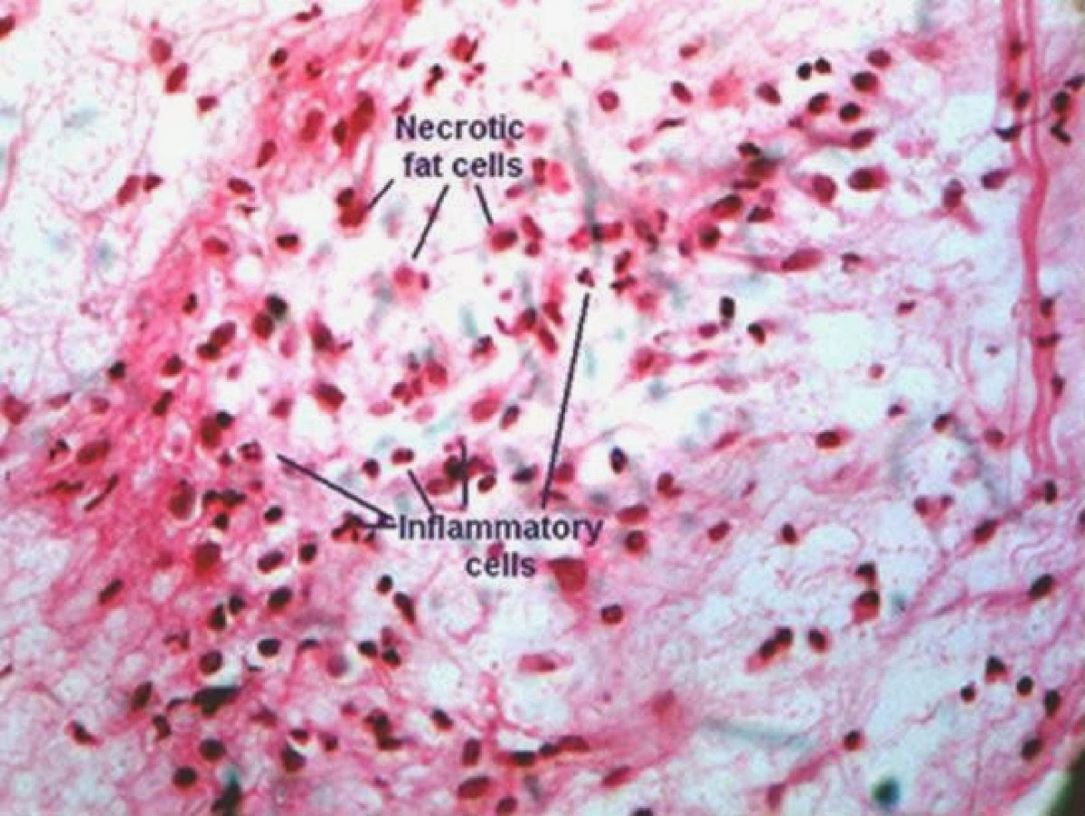
necosisCell death is one of the most crucial events in pathology, affecting every cell type and being a major consequence of ischemia, infection, toxin, and immune reaction. There are two types of a pattern of cell injury and deaths, necrosis and apoptosis. Necrosis refers to a spectrum
of morphologic changes that follow cell death in living organisms. Necrosis is characterized denaturation of cytoplasmic proteins, failure of membrane integrity, break down of cell organelles and swelling of lethally injured cells.


Types of necrosis 

Necrotic cells have different morphological types. It can be classified into a total of five types

Coagulative Necrosis


Coagulative necrosis is the most common type of necrosis in which cells death happen after exogenous or endogenous stimuli, occurring after such stresses as ischemia, virus toxin, physical and chemical injury. It is manifested by severe cell rupture, denaturation, and coagulation of cytoplasmic proteins.
In coagulative necrosis, the texture of the tissue will be normal or firm, but later it may become soft as a result of digestion by macrophages. The myocardial infarction is the typical example of coagulative necrosis. 


Caseous necrosis

Caseous necrosis is a distinctive form of coagulative necrosis in tuberculosis infection. The term caseous is derived from the gross appearance of the area of necrosis. On microscopic examination, the necrotic focus appears as amorphous coarsely granular eosinophilic debris, without any original outline of cells and tissues. This distinctive types of necrosis are generally attributed to the toxic effect of the unusual cell wall of Mycobacterium, which contains complete waxes. 


Liquefactive necrosis


Liquefactive necrosis is a characteristic of focal bacterial or occasionally fungal infections because these agents constitute powerful stimuli to the accumulation of inflammatory cells. The polymorphonuclear leukocytes in the acute inflammatory reactions are endowed with potent hydrolases, capable of completely digesting dead cells. For the reasons of the presence of more abundant lysosomal enzymes or different hydrolases and the lack of any substantial supporting stroma specific to the nerve cells, hypoxic death of cells within the central nervous system often evokes liquefactive necrosis. 


Fibroid necrosis

Fibroid necrosis refers to an alteration of injured tissues which are homogenous, deeply eosinophilic in necrosis. For example, malignant hypertension, the wall of the arterioles is under such necrosis, because of the insulation and accumulation of immunoglobulins, plasma proteins and especially the consequences deposition of fibrin.  

Gangrenous necrosis

Gangrenous necrosis is necrosis putrefaction of the tissues as a result of the action of the saprophytic bacteria, mainly clostridium. There are three types of gangrenous necrosis:
1.       Dry gangrenous
2.       Wet gangrenous
3.       Gas gangrenous 

The primary functions of the thyroid hormone basically include:
Functions of Thyroid Hormone
Heat Production
Increases oxygen consumption and BMK of targeted tissues, especially in liver, cardiac muscles and kidney

Protein Metabolism
Promote the synthesis of proteins and enzymes

Carbohydrate Metabolism
Elevates the glucose level in the blood

Fat Metabolism
Promotes oxidization of fatty acids and strengthen the effects of catecholamine and glycogen of lypolysis.

Effects on growth and development
Essential for mental and physical development in human, especially for the development of brain and bone tissues

Effects on CNS
Increases the effectiveness of permissive antigen and increase the excitability of CNS

Effects of Cardiovascular System

Increases heart rate, cardiac contractility, cardiac output and vasodilatation  

structure of BCR complex
It’s a membrane Ig (IgM), associated glycoporotein, Ig α and Igβ , which has a moderately large cytoplasmic domain. These domains each include a short region important for transmitting a signal indicating antigen has bound. This region is called immune tyrosin based activation motif ( ITAM ).

Functions of BCR complex

On activation by antigen, B cells differentiate into plasma cells producing antibody molecules of the same antigen specificity as the receptors.

The abdominopelvic cavity extends from the diaphragm to the pelvis. It is subdivided into a superior abdominal cavity and aninferior pelvic cavity (Figures 1–9 and 1–10a). The abdominopelvic cavity contains the peritoneal cavity, a potential space lined by a serous membrane known as the peritoneum. The parietal peritoneum lines the inner surface of the body wall. A narrow space containing a small amount of fluid separates the parietal peritoneum from the visceral peritoneum, which covers the enclosed organs. You are probably already aware of the movements of the organs in this cavity. Who has not had at least one embarrassing moment when the contraction of a digestive organ produced a movement of liquid or gas and a gurgling or rumbling sound? The peritoneum allows the organs of the digestive system to slide across one another without damage to themselves or the walls of the cavity.

The abdominal cavity extends from the inferior surface of the diaphragm to the level of the superior margins of the pelvis. This cavity contains the liver, stomach, spleen, small intestine, and most of the large intestine.  The organs are partially or completely enclosed by the peritoneal cavity, much as the heart and lungs are enclosed by the pericardial and pleural cavities, respectively. A few organs, such as the kidneys and pancreas, lie between the peritoneal lining and the muscular wall of the abdominal cavity. Those organs are said to be retroperitoneal (retro, behind). per-i-to_ -NE_ -um per-i-to_ -NE_ -al


The pelvic cavity is the portion of the ventral body cavity inferior to the abdominal cavity. The bones of the pelvis form the walls of the pelvic cavity, and a layer of muscle forms its floor. The pelvic cavity contains the urinary bladder, various reproductive organs, and the distal portion of the large intestine. The pelvic cavity of females, for example, contains the ovaries, uterine tubes, and uterus; in males, it contains the prostate gland and seminal glands. The pelvic cavity also contains the inferior portion of the peritoneal cavity. The peritoneum covers the ovaries and the uterus in females, as well as the superior portion of the urinary bladder in both sexes. Visceral structures such as the urinary bladder and the distal portions of the ureters and large intestine, which extend inferior to the peritoneal cavity, are said to be infraperitoneal.

Priming of Immune system

Normal floraOrganisms of normal flora play an important role in the development of immunologic competence. Animals delivered and raised under completely aseptic conditions have a poorly developed reticuloendothelial system, low serum level of immunoglobulin and none of the antibodies to normal floral antigen that often cross-react with those of pathogenic organisms and confer a degree of protection against them.


Exclusionary Effects

The normal flora produces conditions that tend to block the establishment of e4xtraneous pathogens and their ability to infect the host. The bifidobacteria in the colon of the breastfed infant produce an environment inimical to colonization by enteric pathogens, this protective effect is aided by ingested maternal IgA. Breastfeeding has clearly shown to help to protect infant from enteric bacterial infections.

Production of Essential Nutrients by Normal flora

In humans, member of the vitamin B group and vitamin K are produced by normal flora. Bacterial vitamin production is reduced during broad spectrum antibiotic therapy, and supplementation with vitamin B complex is indicated in malnourished individuals.

The main goals of the respiration are to provide oxygen to the tissues and to remove carbon dioxide. To achieve these goals, respiration can be divided into four major functions. They
  1. Pulmonary Ventilation. Means the inflow and out flow of air between the atmosphere and the lung alveoli.
  2. Diffusion, of oxygen and carbon dioxide between the alveoli and the blood
  3. Transport of oxygen and carbon dioxide in the blood and body fluids to and from the body’s tissue cell
  4. Regulation of ventilation and other focets of respiration
The upper airway consists of the nose, mouth, pharynx, and larynx. The larynx opens into the trachea, which in turn branches into two bronchi, enter into both lungs. The airway beyond larynx can be divided into two zones

Conducting zone
Extends from the top of the trachea to the beginning of the respiratory bronchioles

Functions
Provide low resistance pathway for air flow
Defends against microbes, toxic chemicals, and other foreign matters

Respiratory Zone
Extend from the respiratory bronchioles to the alveoli and is the region where gases exchange with blood. The inner surface of the airway down to the end of the respiratory bronchioles contains cilia, glands and epithelial cells, secrets mucus which keep the lungs clear of particulate matter and bacteria that enters body with dust particles. Another protective mechanism against infection is provided by the macrophages that exist in the airway and alveoli, these macrophages engulf and destroy inhaled particles and bacteria that have reached alveoli.


Reference 
Guyton, A & Hall, J. (2006).Text book of Medical Physiology.11th Edition. Elsevier Saunders